Monday, November 14, 2011

Observation 4- 11/10/11

Hello!

Last Thursday, I observed my MicroAquarium for the last time.  There were some changes from my last observation.  There were still many organisms in the MicroAquarium, but it was not quite as active as last time.   Additionally, there was more activity in the bottom layer of the tank around the silt than ever before.  I also observed two dead organisms at the bottom of the tank.

There will a couple of particularly interesting organisms in my MicroAquarium last Thursday.  One of them was likely a monomatta, which is a type of rotifer that we were unable to find in any texts.


Monommata sp.

Another interesting organism in my MicroAquarium this week was a Philodina.  This one was particularly interesting to me because it was pulling materials toward it and had a very strong current going.  Below, you can see a picture of this Philodina.

Philodina sp.
(Pennak 1989, Figure 3)

 Sources:
http://www.plingfactory.de/Science/Atlas/KennkartenTiere/Rotifers/source/Monommata%20longiseta.html

Pennak, R. W. (1989).  Fresh-water invertebras of the United States: protozoa to mollusca.  New York City, New York: John Wiley and Sons, Inc.

Monday, November 7, 2011

Observation 3- 11/3/2011

I observed my MicroAquarium once again last Thursday, November 3rd.

Many things have changed since my last observation.  For one thing, there were many more organisms this time, including a Euclanis (Pennak 1989, Figure 2), several Vorticella (Patterson 1996, Figure 233), several Actinospaerium (Patterson 1996, Figure 395), a Philodina (Pennak 1989, Figure 3), a Tachysoma (Patterson 1996, Figure 265), and a rotifer that was extremely difficult to identify--our best guess was a Furcularia--(Ward and Whipple 1918, Figure 871).  Below, I will post some of the most interesting of these organisms.


Philodina
(Pennak 1989, Figure 3)

Rotifer-Possibly Furcularia
(Ward and Whipple 1918, Figure 871)
Another change was that Plant B (Utricularia gibba) seemed to be dead/dying.  Below, there is a picture of the dead plant material.  This plant material was rich with organisms feeding on the remaining matter.

Dead Plant Material -  Utricularia gibba 
 This week was the first week in which I observed any dead organisms on the bottom of the tank.  Below, you can see a picture of that organism, which I was unable to identify.  You can't tell in the picture, but the organism was a hive of activity for other organisms, which seemed to be feeding on the dead organism.
Unidentified Dead Organism

I plan to observe my MicroAquarium again later this week and update this blog with more information soon after.


Sources:

Bick, H. (1972).  Ciliated protozoa.  Geneva, Switzerland: World Health Organization.

Patterson, D.J. (1996).  Free-living freshwater protozoa: a colour guide.  Washington, D.C.: ASM Press.

Pennak, R. W. (1989).  Fresh-water invertebras of the United States: protozoa to mollusca.  New York City, New York: John Wiley and Sons, Inc.

Ward, H. B. and Whipple, G. C. (1918).  Fresh-water biology.  New York City, New York: John Wiley and Sons, Inc.

Monday, October 31, 2011

Observation 2-10/27/11

I observed my MicroAquarium for the second time last Thursday, the 27th of October.

Earlier in the week, Dr. McFarland added a food pellet to the MicroAquarium. On Friday, October 21, 2011, one Beta Food Pellet was inserted into the MicroAquarium ("Atison's Betta Food" made by Ocean Nutrition, Aqua Pet Americas, 3528 West 500 South, Salt Lake City, UT 84104).  The ingredients in this beta pellet were fish meal, wheat flower, soy meal, krill meal, minerals, vitamins and preservatives and the analysis of this shows the following: Crude Protein 36%, Crude fat 4.5%, Crude Fiber 3.5%, Moisture 8%, and Ash 15%.

This week I observed many of the same organisms that I observed last week.  I saw a nematoda, a Cyclopoid nauplius, a tachysoma, and several unidentified organisms once again.  Many of these organisms were in the middle layer of the MicroAquarium.  They were also not quite as active as last time I observed the habitat.  The Cyclopoid naupilius was particularly sluggish this time when compared to the speed of its movement during Observation 1 on October 20th.


I would also like to note that the area around Plant B (Utricularia gibba L. flowering plant) seemed to be much more active than the area around Plant A (Amblestegium sp. Moss ).


I plan to observe my MicroAquarium again later this week.


Monday, October 24, 2011

Observation 1-10/20/11

I observed my MicroAquarium on October 20, 2011 (approximately 9 days after putting it together).  To recap, my water and substrate was from Source 8 (the Tennessee River near Neyland Stadium), and I added moss from  a natural spring at Carters Mill Park (Amblestegium sp.) and whose original material was from south shore of Spain Lake ( in White County and grown in water tanks outside of greenhouse at Hesler Biology Building (ricularia gibba L.).


During this observation, I noted several organisms in the water and among the mosses.  I did not, however, find any organisms in the substrate.


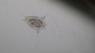
The first organism I saw I identified as Actinosphaerium (Patterson 1996, Figure 395).  I saw several of these organisms throughout the MicroAquarium but mostly in the middle and lower levels.  Below is a picture of one such organism.


Actinospaerium
(Patterson 1996, Figure 395)
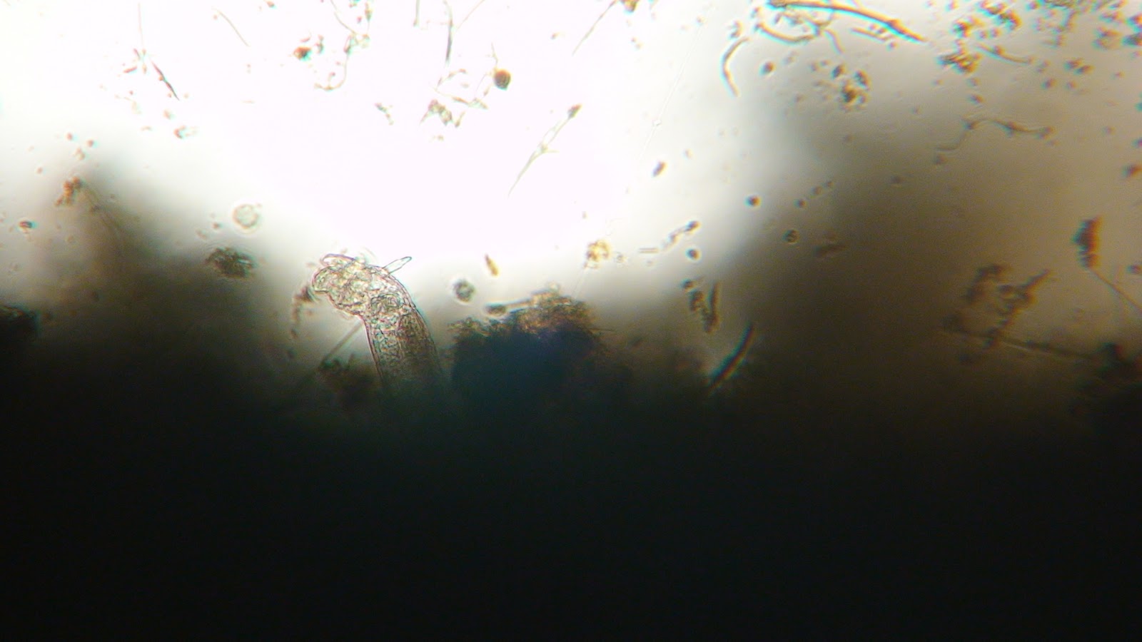
I also identified Nematoda (Smith 2001, Figure 8.1), which appeared to be stuck in one of the moss samples.  This Nematoda, which I could not classify more specifically, can be seen in the picture below.

Nematoda
 (Smith 2001, Figure 8.1)

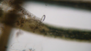
Additionally, I identified the nymph of a Cyclpoid nauplius (Smith 2001, Figure 19.6).  This organism was very fast-moving and hard to capture on camera.  An image of the immature copepoda can be seen below.

Cyclopoid nauplius Nypmh
(Smith 2001, Figure 19.6)
Below is an image of Vorticella (Patterson 1996, Figure 233).  This organsim could be found in many areas of the MicroAquarium.  The movement of the organism is interesting in that it pulls water through itself in order to eat.  Additionally, I witnessed it curling up when it was hit by a larger organism that was careening around the habitat (classification of larger organism unknown).

Vorticella
(Patterson 1996, Figure 233)

I was also lucky enough to have  a protozoa called Campanella in my MicroAquarium (Bick 2972, Figure 61).  To me, this organism looked like a two-headed vorticella and appeared to function similarly (pulling water through itself to catch organisms).

Campanella
(Bick 2972, Figure 61)

I saw several other organisms in my MicroAquarium, including a Tachysoma (Patterson 1996, Figure 265), several kinds of algae, and several unidentified organisms.  I plan to observe my MicroAquarium again on Thursday.

Sources:
Bick, H. (1972).  Ciliated protozoa.  Geneva, Switzerland: World Health Organization.

Patterson, D.J. (1996).  Free-living freshwater protozoa: a colour guide.  Washington, D.C.: ASM Press.

Smith, D. G. (2001).  Pennak's freshwater invertebrates of the United States.  New York, NY: John Wiley 
and Sons, Inc.


Monday, October 17, 2011

Week 1: Setting Up My MicroAquarium (10/11/11)

Hello!


This past week in lab, we began our MicroAquarium project.  I will now outline the procedure I followed to set up the aquarium.


First, I obtained a glass tank, stand holder, and lid.  Then I put color dots to mark the tank as mine (dark blue, yellow, and green with my initials written on them).  I placed these dots in the top left hand corner.


Using a pipet, I extracted water from sample number 8, which contained water from the Tennessee River at the boat ramp across from the Knoxville sewer plant (Neyland Dr. Knox Co. Knoxville TN. Full sun exposure. French Broad and Holston Rivers water Sheds N35 56.722 W83 55.587 813 ft 10/9/2011).  I extracted enough water from the bottom of the container to fill the MicroAquarium until it was about 1/3 full, the next 1/3 of the water I extracted from the middle layer of the water, and I filled the rest of the MicroAquarium tank with water from the surface of the water.  Then I placed it in the stand and inserted two types of plants.  The first plant was Plant A or Amblestegium sp. Moss collected from a natural spring at Carters Mill Park (Carter Mill Road, Knox County, TN N36 01.168 W83.42.832) in partial shade exposure obtained on 10/9/2011.  The next plant I added was Plant B or Utricularia gibba L. flowering plant (a carnivorous plant) whose original material was from south shore of Spain Lake (N 35degrees55 12.35" W08degrees20' 47.00), Camp Bella Air Rd East of Sparta Tennessee in White County and grown in water tanks outside of greenhouse at Hesler Biology Building (The University of Tennessee, Knox County, Knoxville, TN).


After setting all of this up, I observed the contents of the MicroAquarium under the microscope for the first time.  I did not see any moving organisms.  The plants were chlorophyll green.  Organisms had not yet settled well enough after the move to be able to ascertain which layered they preferred (top, middle, or bottom). 


I plan to observe my MicroAquarium for the second time on Thursday, October 20th and will update with more information then.

Tuesday, September 27, 2011

Testing

I was required to make this blog for a botany class I'm taking. I am now testing it out. It seems to work okay.